Exploring the limits to quantitative elastography: supersonic shear imaging in stretched soft strips
 model vs. measurement
Abstract
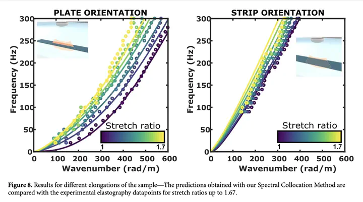
Objective. Shear wave elastography has enriched ultrasound medical imaging with quantitative tissue stiffness measurements. We aim to explore the limitations that persist related to viscoelasticity, guiding geometry or static deformation. Approach. A nearly-incompressible soft elastomer strip is chosen to mimic the mechanical behaviour of an elongated tissue. A supersonic shear wave scanner measures the propagation of shear waves within the strip. It provides a wide range of shear wave velocities, from 2 to 6 m s−1, depending on the frequency, the static strain as well as the orientation of the strip. Main results. To explain these different measurements, the guided wave effect is highlighted and analysed from the dispersion diagrams provided by the spatio-temporal Fourier transform of the raw data. The guided waves are then described using a material model that accounts for both the rheology and the hyperelastic behaviour, and allows to extract the mechanical parameters of the sample. Significance. To overcome some limitations of current elastography, we propose a theoretical framework which allows the simultaneous characterization of the viscoelastic and hyperelastic properties of soft tissues, paving the way for robust quantitative elastography of elongated tissues.
Daniel A. Kiefer
Researcher at Institut Langevin
Research in guided elastodynamic waves, fluid-structure interaction, simulation and signal processing for ultrasonic sensors and nondestructive testing.